Dental implants rely on precision, not guesswork. A CBCT scan for dental implants provides implant dentists in Ballwin, MO with a detailed 3D view of the jaw, allowing safer planning, accurate placement, and more predictable long-term results. By replacing estimates with precise imaging, CBCT scans support guided implant surgery that protects vital structures and improves implant stability from the start.
If you are researching dental implants in Ballwin or searching for dental implants near me, understanding how implant planning works can help you make confident decisions. At ActOn Implants, advanced imaging is used early in the process so each implant plan is built around your anatomy, not averages. Scheduling a consultation that includes CBCT imaging is often the first step toward a treatment plan designed to last.
Why Accurate Implant Planning Matters for Long-Term Implant Success
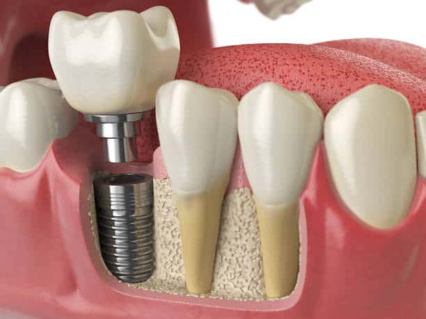
Accurate implant planning also determines how chewing forces move through the implant and into the surrounding bone. When you bite or chew, pressure travels from the crown down through the implant and into the jaw. Natural teeth are supported by a periodontal ligament that helps absorb and distribute this force. Dental implants do not have this cushioning layer, which means force transfer is more direct and placement accuracy becomes critical.
If an implant is angled improperly or placed in a less supportive area of bone, pressure may concentrate in specific spots instead of being evenly distributed. Over time, this can place added stress on the surrounding bone, the implant itself, and the restoration attached to it. Uneven force can contribute to bone loss around the implant, loosening of components, or premature wear of the crown, even if the implant initially feels stable.
This is why implant position matters beyond what appears on a standard X-ray. Two implants may look similar in a flat image, yet function very differently years later. For example, an implant placed slightly off-axis may handle normal chewing at first but experience higher stress during daily use. Another implant, positioned with proper angulation and bone support, can distribute force more evenly and remain comfortable and stable over the long term.
Careful planning helps ensure implants are placed where bone can best support functional forces, not just where space appears available. By accounting for how pressure moves through the implant system from the start, accurate planning supports durability, comfort, and long-term implant success.
The Risks of Placing Dental Implants Without Advanced Imaging
Implant complications are often tied to planning limitations rather than the implant itself. Traditional imaging does not always show depth, bone quality, or the exact location of nerves and sinuses.
Potential issues associated with limited imaging include:
- Nerve irritation or numbness due to implant proximity
- Sinus complications during upper jaw placement
- Weak bone support that affects implant stability
- Increased long-term implant failure risk
CBCT scans help reduce these concerns by allowing implants to be planned around each patient’s unique anatomy instead of relying on estimates during surgery.
What Is a CBCT Scan and How Does It Work for Dental Implants?
A CBCT scan, or cone beam computed tomography scan, creates a three-dimensional image of the jaw that functions like a detailed map rather than a flat picture. For dental implants, this implant diagnostic imaging allows dentists to evaluate bone structure, density, nerves, and sinuses before any surgical steps take place.
This level of detail supports decisions about implant size, angle, and placement location. Instead of discovering challenges during surgery, CBCT for dental implants allows those factors to be addressed during planning, leading to smoother procedures and more predictable outcomes.
CBCT vs Traditional Dental X-Rays for Implant Planning
Traditional dental X-rays remain useful, but they show limited information when implants are involved. The difference becomes clear when comparing 2D and 3D dental imaging.
| Imaging Method | Traditional Dental X-Rays (2D) | CBCT Scans (3D) |
|---|---|---|
| Bone visualization | Show height but not depth of bone | Show bone height, width, and density |
| Nerve and sinus visibility | Limited nerve and sinus visibility | Clear mapping of nerves and sinuses |
| Implant angulation | Less precise implant angulation | Accurate angle and depth planning |
| Planning confidence | Higher reliance on estimates | Confident, guided implant placement |
CBCT imaging is preferred for implant dentistry because it replaces guesswork with measurable detail, improving both safety and accuracy.
How CBCT Improves Implant Precision in Ballwin, MO
CBCT scans play an active role in how implants are placed, not just how they are evaluated. For Ballwin patients, this means implant plans that account for bone quality, anatomical limitations, and long-term function before surgery begins. The result is a more controlled process with fewer surprises.
This level of planning is especially valuable for patients seeking long-term solutions rather than short-term fixes. When implant placement is mapped carefully, outcomes tend to feel more natural and stable over time.
Mapping Bone Density and Bone Volume Before Implant Placement
Bone density and volume play a major role in how well an implant can anchor and remain stable over time. CBCT imaging allows dentists to evaluate bone density for implants, helping determine whether the jaw can provide reliable implant bone support before placement. This level of detail is especially important when assessing areas that may have weakened after tooth loss.
With CBCT scans, dentists can assess:
- Bone thickness and height
- Areas of insufficient bone for implants caused by previous extractions or bone loss
- Bone quality needed to support daily chewing forces
For example, thinner or less dense bone may require a different implant size, placement angle, or additional preparation compared to denser bone. Identifying these factors early allows the treatment plan to be adjusted before surgery, supporting stronger initial stability and long-term implant success.
Identifying Nerves, Sinuses, and Anatomical Limitations
Nerves and sinus spaces vary from person to person, particularly in the upper jaw. CBCT scans clearly show where these structures are located so implants can be placed safely. This planning helps avoid post-treatment numbness, sinus complications, or discomfort related to implant positioning.
By addressing these considerations in advance, CBCT imaging allows implant dentists to proceed with confidence and gives patients peace of mind throughout the process.
CBCT and Guided Implant Surgery: How They Work Together
CBCT imaging and guided dental implant surgery work together to improve accuracy from planning through placement. The CBCT scan provides the detailed data needed for digital implant planning, allowing implant position to be mapped before surgery begins. This planning-first approach improves efficiency during the procedure and supports higher implant precision that Ballwin patients can rely on for long-term results.
The guided implant process typically follows these steps:
- A CBCT scan captures detailed 3D images of the jaw
- Digital planning software determines the ideal implant position
- Virtual placement is reviewed and refined before surgery
- A custom surgical guide is created based on the plan
- Implants are placed using the guide to control angle and depth
This approach allows implants to be placed according to a verified plan rather than adjusted during surgery. Patients who want a clearer understanding of how accuracy is achieved often explore guided implant options during their consultation.
Digital Implant Planning and Surgical Guides
Digital implant planning improves consistency by allowing each step to be evaluated and refined before surgery takes place. Surgical guides created from CBCT data help control implant placement during the procedure, reducing variability and improving predictability. This planning process supports precise execution, especially in cases where accuracy directly affects comfort and longevity.
At ActOn Implants, planning, placement, and restoration are completed under one roof, which supports continuity and efficiency. For patients seeking a more precise approach, guided dental implant surgery paired with CBCT imaging offers a clear advantage in achieving stable, long-lasting results.
Who Benefits Most from CBCT-Based Implant Planning?
CBCT-based planning benefits all implant patients, but it becomes especially valuable in cases where anatomy, bone quality, or treatment complexity increases the margin for error. In these situations, detailed imaging helps guide decisions that directly affect implant stability, comfort, and longevity.
Patients with Bone Loss or Previous Tooth Extractions
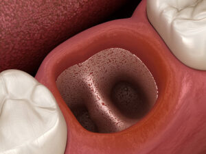
After a tooth is lost, the surrounding bone naturally begins to shrink. Without CBCT imaging, it can be difficult to determine how much bone remains or whether it can reliably support an implant. Planning based only on surface appearance or limited imaging may lead to implants being placed in weaker bone areas.
CBCT scans allow dentists to see exactly where bone has resorbed and where stronger support still exists. This information helps determine whether bone grafting is necessary, whether implant size or position should be adjusted, or whether an alternative approach will provide better long-term stability. For patients with prior extractions, CBCT planning helps ensure implants are placed intentionally, not reactively.
Full-Arch and Multiple-Tooth Implant Cases
Replacing multiple teeth requires careful coordination so implants work together as a system rather than independently. Without detailed planning, implants may be spaced unevenly or placed at angles that concentrate biting forces instead of distributing them evenly.
CBCT imaging supports precise alignment and spacing by showing bone contours and density across the entire arch. This allows implants to be positioned where they can best support chewing forces over time. For full-arch cases, this planning helps improve comfort, reduce mechanical stress on restorations, and support long-term function rather than short-term stability alone.
Patients Considering Implant-Supported Dentures
Implant-supported dentures rely on strategic implant placement to achieve stability and minimize movement. Without CBCT imaging, implant positioning may be based on estimates rather than exact bone availability, which can affect how securely the denture fits.
CBCT scans help identify the strongest bone regions and guide implant placement to maximize support. This planning improves denture stability, reduces pressure points, and helps create a more secure, natural-feeling result. For patients transitioning from removable dentures, CBCT-based planning plays a key role in achieving a noticeable improvement in comfort and confidence.
What Ballwin Patients Can Expect During a CBCT Scan
A CBCT scan is fast, non-invasive, and designed for patient comfort. The scan itself typically takes less than a minute and does not involve needles, injections, or physical contact. CBCT imaging also uses a focused, low-dose radiation field tailored for dental diagnostics, making it appropriate for implant planning when detailed information is needed.
Beyond the scan itself, the most important part of the CBCT process happens after the images are captured. The scan gives implant dentists a detailed, three-dimensional view that can be reviewed from multiple angles, allowing them to evaluate bone thickness, bone density, and the exact location of nearby anatomical structures. This information helps transform a general treatment idea into a precise, personalized implant plan.
For patients, this planning step often answers important questions early in the consultation process. CBCT imaging helps clarify whether existing bone can support an implant, if grafting is needed, or if placement should be adjusted to improve long-term stability. Instead of discovering these details during surgery, they are addressed upfront, which helps reduce uncertainty and avoid mid-procedure changes.
CBCT scans also support clearer communication between the dentist and patient. Because the images are three-dimensional, patients can often see why a particular recommendation is made. This makes it easier to understand treatment options, timelines, and expected outcomes, helping Ballwin patients move forward with fewer surprises and greater confidence.
Schedule a Dental Implant Consultation in Ballwin, MO
Dental implant planning is most effective when advanced imaging is part of the evaluation. CBCT scans allow implant dentists to create personalized treatment plans based on your anatomy rather than assumptions.
At ActOn Implants, implant planning, surgery, and restoration are handled in one location for consistency and efficiency. If you are looking for an implant dentist near me or want to understand your options for dental implants in Ballwin, MO, a consultation that includes CBCT imaging can help clarify the path forward.
FAQs About CBCT and Dental Implants
Why is CBCT important in implants?
CBCT is important because it provides a three-dimensional view of the jaw that supports accurate implant placement. This imaging helps dentists identify bone quality, nerve location, and anatomical limitations before surgery. The result is safer placement and more predictable outcomes.
What are the benefits of CBCT in dentistry?
CBCT imaging improves diagnostic accuracy and supports detailed treatment planning. In implant dentistry, it enhances precision, reduces complication risk, and helps dentists explain treatment options more clearly to patients.
What is the role of CBCT in presurgical planning of dental implants?
CBCT allows dentists to plan implant size, position, and angle digitally before surgery. This planning supports guided implant surgery and reduces the need for adjustments during the procedure, leading to smoother treatment and consistent results.
How to improve dental implant success?
Implant success improves with accurate imaging, careful planning, and guided placement techniques. CBCT scans support each of these factors by providing detailed anatomical information that informs better decisions from the start.